Cuboid Bone Siding

Cuboid Bone Wikipedia

Cuboid Bone Os Cuboideum Bones Cuboid Lion Sculpture

The Bones In The Foot Inferior View Picture Illustrated From Thieme Atlas Of Anatomy General Anatomy And Musculoskel Foot Anatomy Body Anatomy Anatomy Bones

Human Muscle Quiz Printout Of Foot Anatomy Bones Skeletal System Joints Of Foot Muscles Anatomy Bones Foot Anatomy Anatomy

Middle Cuneiform Bone Medical Anatomy Human Anatomy And Physiology Human Anatomy

Cuboid Bone An Overview Sciencedirect Topics

The cuboid bone is located between the base of the foot and the ankle.
Cuboid bone siding.

Skeleton Feet Foot Bones Top View Your Foot Is Quite Easy To Find Just Look Down Anatomy Bones Foot Anatomy Foot Anatomy Bones

Bones In The Foot Powerpoint Diagram Diagram Relationship Diagram Feet

Diagram Of Hand And Wrist Wrist Hand Anatomy Bones Wrist Anatomy Medical Anatomy

The Foot Anatomy Foot Anatomy Anatomy Bones Ankle Anatomy

Mnemonic For Learning Tarsal Bones Human Anatomy And Physiology Medical Mnemonics How To Memorize Things

A Longitudinal Section Of A Femur Bone Showing Long Bone Structure Human Body Bones Anatomy Bones Human Bones

Bones Of The Foot Coloring Bone Color Anatomy Color

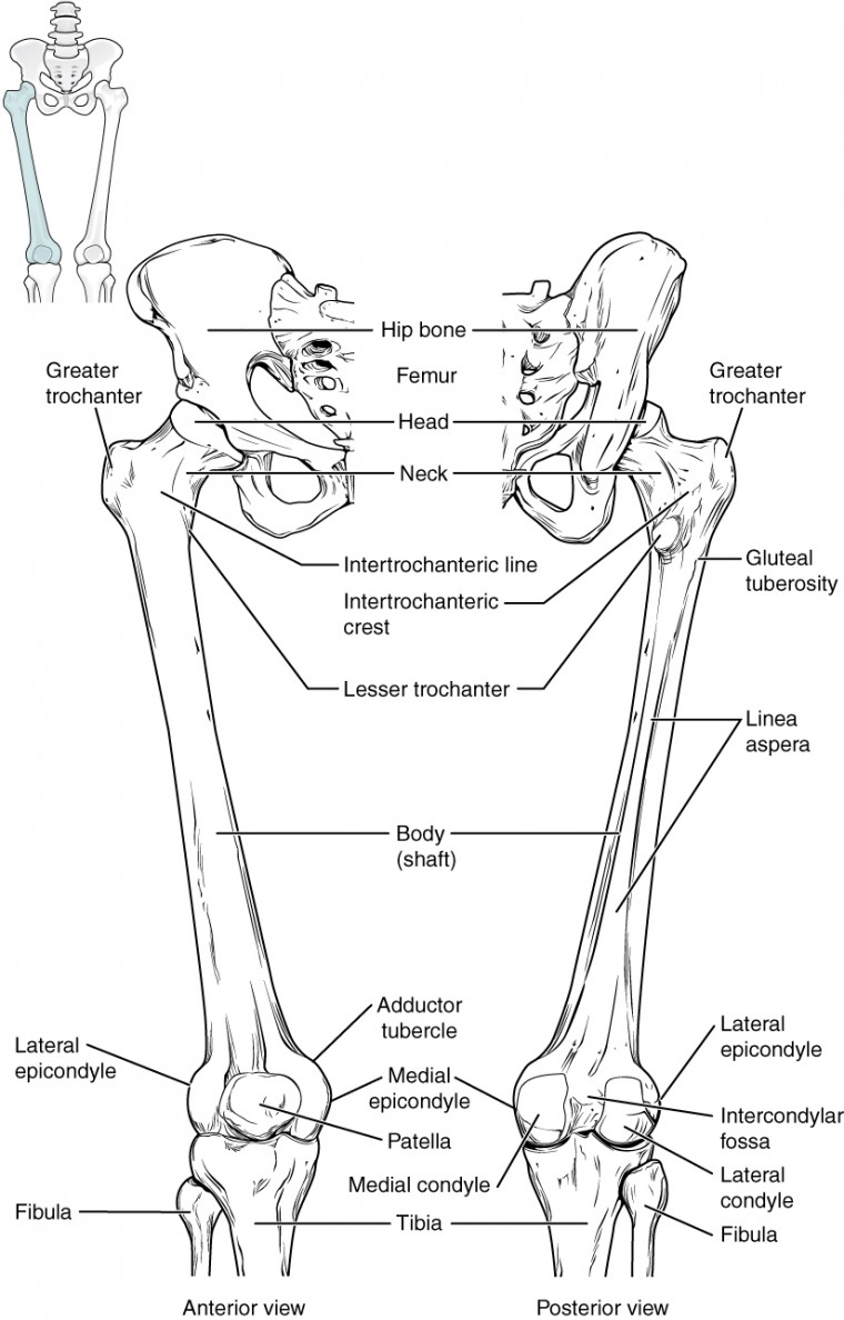
The Femur Consists Of Four Parts The Head Greater Trochanter Lesser Trochanter Anatomy Bones Physiology Human Anatomy
Bones Of The Lower Limb Anatomy And Physiology

Anterior And Posterior View Of The Radial And Ulnar Bones Human Anatomy And Physiology Anatomy Bones Anatomy And Physiology

Tibia Fibula Anatomy Tibia And Fibula Diagram Google Search Anatomy 1 Pinterest Anatomy Bones Human Bones Anatomy Human Anatomy And Physiology

Osteology Of The Skull Anatomy 502 Shows Optic Groove In 2020 Osteology Anatomy Bones Skull Anatomy

Thoracic Cage Is Made Up Of Bones And Cartilage Along It Consists Of The 12 Pairs Of Ribs With Their Costal Cartilages Thoracic Cage Anatomy Bones Human Bones
Ankle Ligaments Netter Ankle Anatomy Foot Anatomy Muscle Anatomy

Foot Care Human Anatomy And Physiology Feet Care Anatomy Bones

Www2 Highlands Edu Academics Divisions Scipe Biology Faculty Harnden 2121 Images Scapula Jpg Anatomy Bones Human Anatomy And Physiology Anatomy

Short Bones Anatomy And Physiology Gross Anatomy Pearson Education

Ankle Bones Diagram Koibana Info Ankle Anatomy Foot Anatomy Human Anatomy
Https Encrypted Tbn0 Gstatic Com Images Q Tbn 3aand9gctdm5f3xswfvxw9 Hdzwr0jp0gqsfifzgludrj9nkednrmjzrzq Usqp Cau

Here Are The Superior And Inferior Views Of The Food Bones Skeletal System Nursing Degree System

This Image Depicts The Shoes Of Bones Examples Of Flat Bones Are The Scapulas And The Sternum Examples Of Irregular Bones Are The Sphenoid Bone Scapula Image

Pin On Mss

Pin By Genna Hornsby On Anatomy Human Anatomy And Physiology Anatomy Bones Medical Anatomy

Pin By Holly Laws On Skeleton Project Types Of Bones Bones And Muscles Bones
Source : pinterest.com
